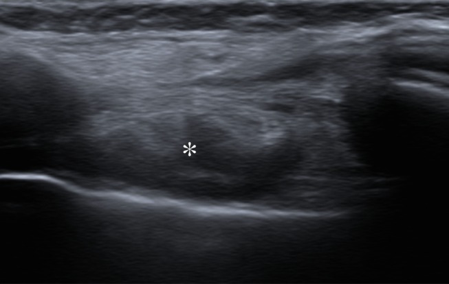
Figure 1d:

Representative case of a 47-year-old woman after left mastectomy and silicone implant reconstruction 6 years earlier. IMLN was interpreted as suspicious for metastatic disease, and consequently the patient underwent full oncologic work-up. (a) Axial silicone-suppressed implant-protocol MR image of the left reconstructed breast demonstrates an enlarged left IMLN (*). (b) PET/CT was performed because of concern for metastatic disease, and the left IMLN (*) was FDG avid on the fused attenuated corrected image. (c) The CT component of the same PET scan demonstrates the left IMLN (*). (d) Targeted ultrasonography (US) was then performed; it depicted the left IMLN, which was amenable to percutaneous biopsy. Pathologic evaluation showed marked reactive change with dense histiocytic reaction and numerous lipophages consistent with a benign cause (*).
