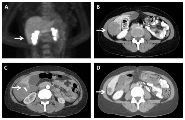
Fig. 1.
Images illustrating (A) the patient’s initial positron emission tomography (PET) scan for staging without evidence of hepatic metastasis (arrow indicating future area of metastasis), and computed tomography (CT) illustrating (B) the patient’s liver metastasis (arrow) prior to initiation of treatment, (C) active extravasation of contrast from the tumor (arrow) after three doses of combined targeted therapy, and (D) durable response (arrow) with re-initiation of therapy after resolution of the hemorrhagic event.

