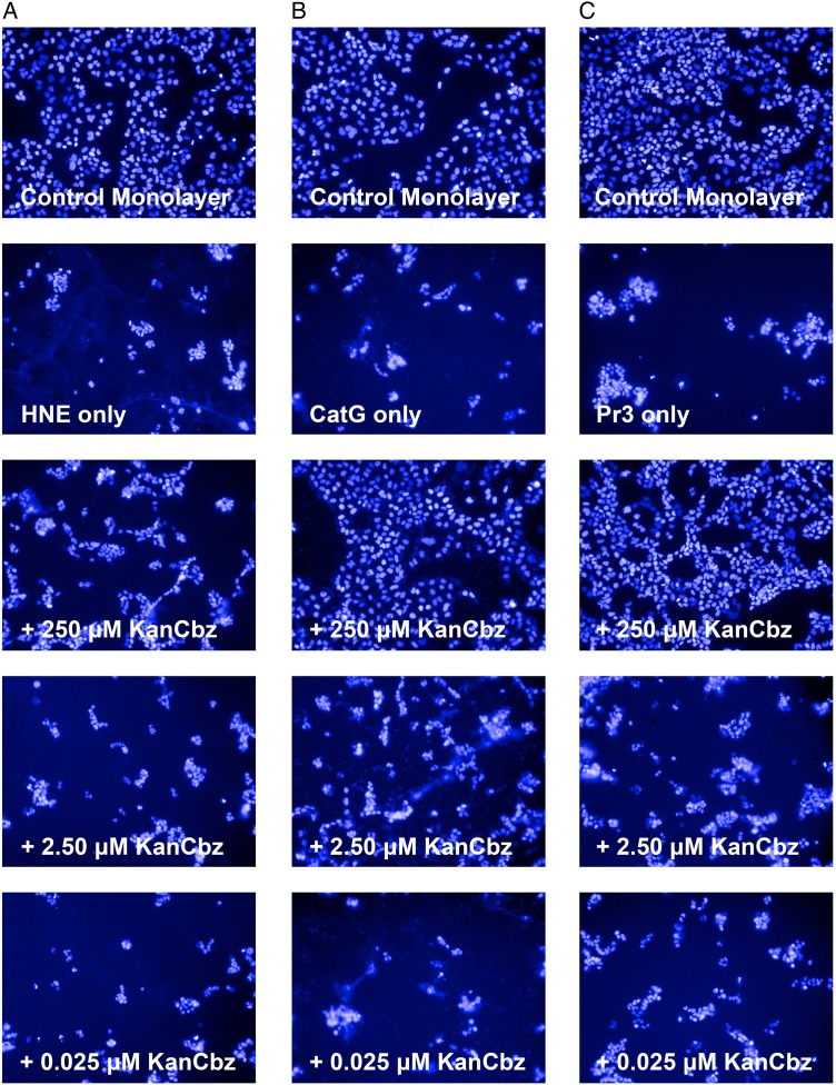
Fig. 4.
Protection against protease-mediated cell detachment. A549 lung epithelial cells were exposed to (A) HNE (50 nM), (B) CatG (250 nM) or (C) Pr3 (50 nM) in the presence or absence of decreasing concentrations of KanCbz. The NSPs were pre-incubated with KanCbz (250, 2.50 and 0.025 μM) for 30 min on ice before addition to confluent A549 cells. After 24 h lung epithelial cell nuclei were stained with Hoechst 33342 and cells were imaged using Operetta High Content Imaging System. Only one representative field for each experiment is shown for clarity. At 250 μM KanCbz inhibits HNE-, CatG- and Pr3-induced cell detachment. At lower concentrations, this protection is lost and protease-mediated cell detachment is once again observed. This figure is available in black and white in print and in color at Glycobiology online.

