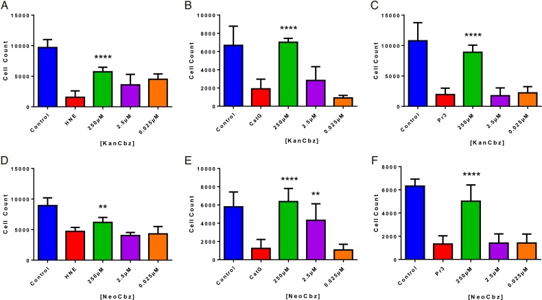
Fig. 5.
Quantification of NSP-mediated cell detachment in the presence of two lead compounds. A549 lung epithelial cells were exposed to each NSP (50 nM HNE, 250 nM CatG or 50 nM Pr3) in the presence of decreasing concentrations of KanCbz (A–C) or NeoCbz (D–F). After 24 h lung epithelial cell nuclei were stained with Hoechst 33342 and cells were imaged and counted using an Operetta High Content Imaging System and Harmony Analysis Software, respectively. At the highest concentration, both KanCbz and NeoCbz protected cells against protease-mediated cell detachment. Data are presented as mean + SE, **P < 0.01, ***P < 0.001 ****P < 0.0001 when compared with protease-treated cells (n = 3 from three experiments each done in triplicate). This figure is available in black and white in print and in color at Glycobiology online.

