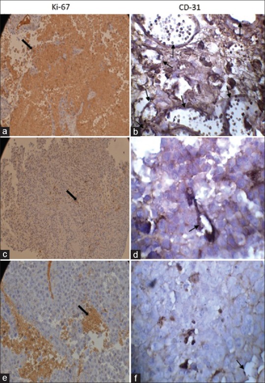
Figure 2.

Immunostaining of CD31 and Ki-67 in melanoma tumor, (a and b) leptin, (c and d) 9F8, (e and f) control groups. Leptin treated animals appeared to have significantly higher percentage of CD31 and Ki-67 staining in their tumors while no significant difference was found between other groups (*P <0.05). Original magnification ×400. Note the strong brown staining in tumors excised from leptin treated mice. Arrows depict cells in tumor with intensive staining
