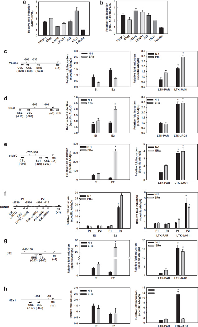
Figure 1.
Active Notch-1 facilitates the transcription of ERα-target promoters in the absence of E2. In all experiments, MCF-7 cells were grown in phenol red-free RPMI containing 10% DCC-fetal bovine serum for 3 days prior to harvest. (a) MCF-7 cells were transiently transfected with the active form of Notch-1 (NIC) or pcDNA vector control. The mRNA levels of VEGFα, CD44, c-MYC, CCND1, pS2, HEY1 and β-tubulin were measured by real-time RT–PCR after 48 h after transfection. Values are expressed as relative fold induction by NIC over pcDNA, after internal normalization for 18S rRNA. (b) MCF-7 cells were co-cultured with mouse fibroblasts expressing Jagged-1 (LTK–JAG1) or vector-transfected controls (LTK–PAR) for 12 h prior to harvest. The mRNA levels of VEGFα, CD44, c-MYC, CCND1, pS2, HEY1 and β-tubulin were measured by real-time RT–PCR using validated human-specific primers. Values are expressed as relative fold induction by LTK–JAG1 over LTK–PAR, after internal normalization for RPL13a mRNA. (c–h) The schematics of the indicated promoters and ChIP assays. Charcoal-stripped MCF-7 cells were treated with 5 nm E2 or ethanol (vehicle) for 1 h (left), or co-cultured with LTK cells for 3 h (right) before formaldehyde fixation. ChIP assays were performed with antibodies to Notch-1 or ERα, followed by real-time PCR analysis of the indicated regions of each promoter (arrows). Values are expressed as relative fold increase of specific antibody pull-down over IgG control, after normalization for internal control RPL13a. tts, transcription start site; *P < 0.001. ChIP, chromatin immunoprecipitation; ERα, estrogen receptor-α; LTK–JAG1, mouse LTK fibroblasts expressing Notched ligand Jagged-1; LTK–PAR, control vector transduced parental fibroblast; NIC, active form of Notch-1 (Notch-1IC); RPMI, Rosewell Park Memorial Institute; RT–PCR, reverse transcription–PCR; VEGF, vascular endothelial growth factor.

