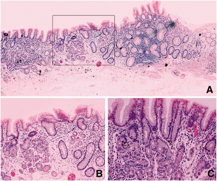
Figure 3.
Histology (hematoxylin & eosin staining) showing heterotopic gastric mucosa of pyloric type in the rectum: (A) seriated section (magnification x5); (B) magnified view of border between gastric and rectal epithelium (boxed area in figure 3a) (magnification x10); C) predominant pyloric mucous glands with rare parietal (P) and endocrine (E) cells (magnification x20).

