
Figure 6. Axotomy-induced activation of phagolysosomes in engulfing glia.
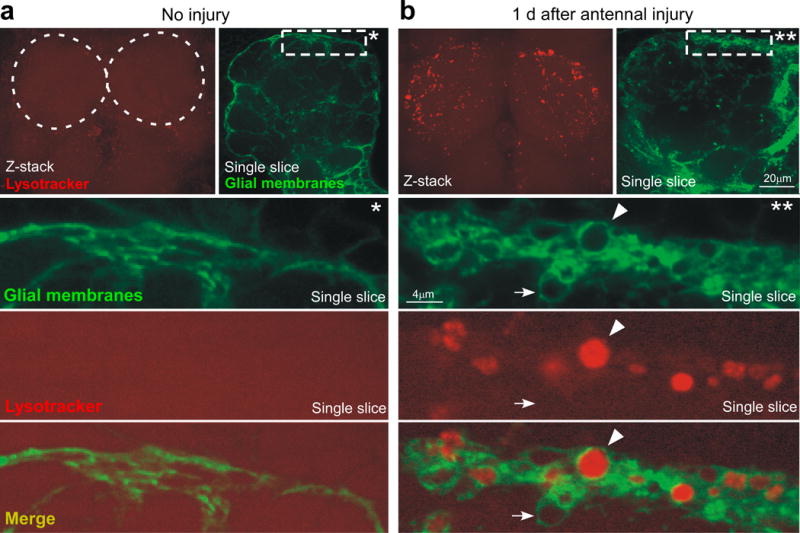
(a) Uninjured animals (w;UAS-mcd8::GFP/+;TIFR-Gal4/+) were stained for Lysotracker Red (confocal Z-stack, top) and visualized for GFP+ glial membranes (single confocal slice, top). Dotted circles; location of antennal lobes. *; high magnification view of the marked rectangle in a single confocal section.
(b) Animals (w;UAS-mcd8::GFP/+;TIFR-Gal4/+) shown 1 day after bilateral antennal injury were stained for Lysotracker Red (Z-stack top) and visualized for GFP+ glial membranes (single slice, top). **; high magnification view of the marked rectangle in a single confocal section. Arrow; an injury-induced glial vesicle. Arrowhead; glial vesicle positive for lysosomal activity.