Figure 1.
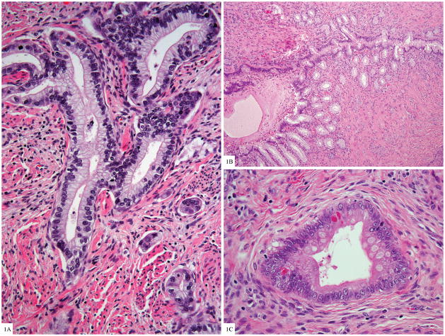
Figure 1A. Gastric type endocervical adenocarcinoma with voluminous clear cytoplasm and basally located nuclei with moderate cytologic atypia, adjacent small invasive tumor clusters (H&E 100×).
Figure 1B. Lobular endocervical glandular hyperplasia with central dilated gland and smaller glands proliferating along the periphery, similar to pancreatic ducts (H&E 40×)
Figure 1C. Goblet cell and neurosecretory-like granules in glands of gastric type adenocarcinoma (H&E 200×)

