Figure 1.
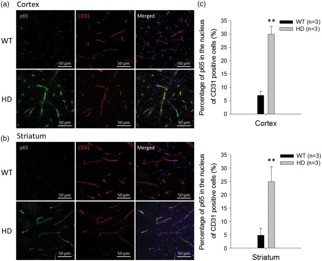
Aberrant NF-κB p65 signaling in brain capillaries of R6/2 HD mice and WT controls. The nuclear distribution of the p65 subunit of NF-κB in the cortex (a) and striatum (b) were identified by immunostaining p65 (green) and CD31 (red) in brain capillaries. Nuclei were stained with Hoechst 33258 (blue). (c) The percentages of p65 in the nuclei of CD31-positive capillary endothelial cells were quantified from immunostaining images. Data are presented as the mean ± SEM of three animals. Scale bars indicate 50 µm. (**P < 0.01).

