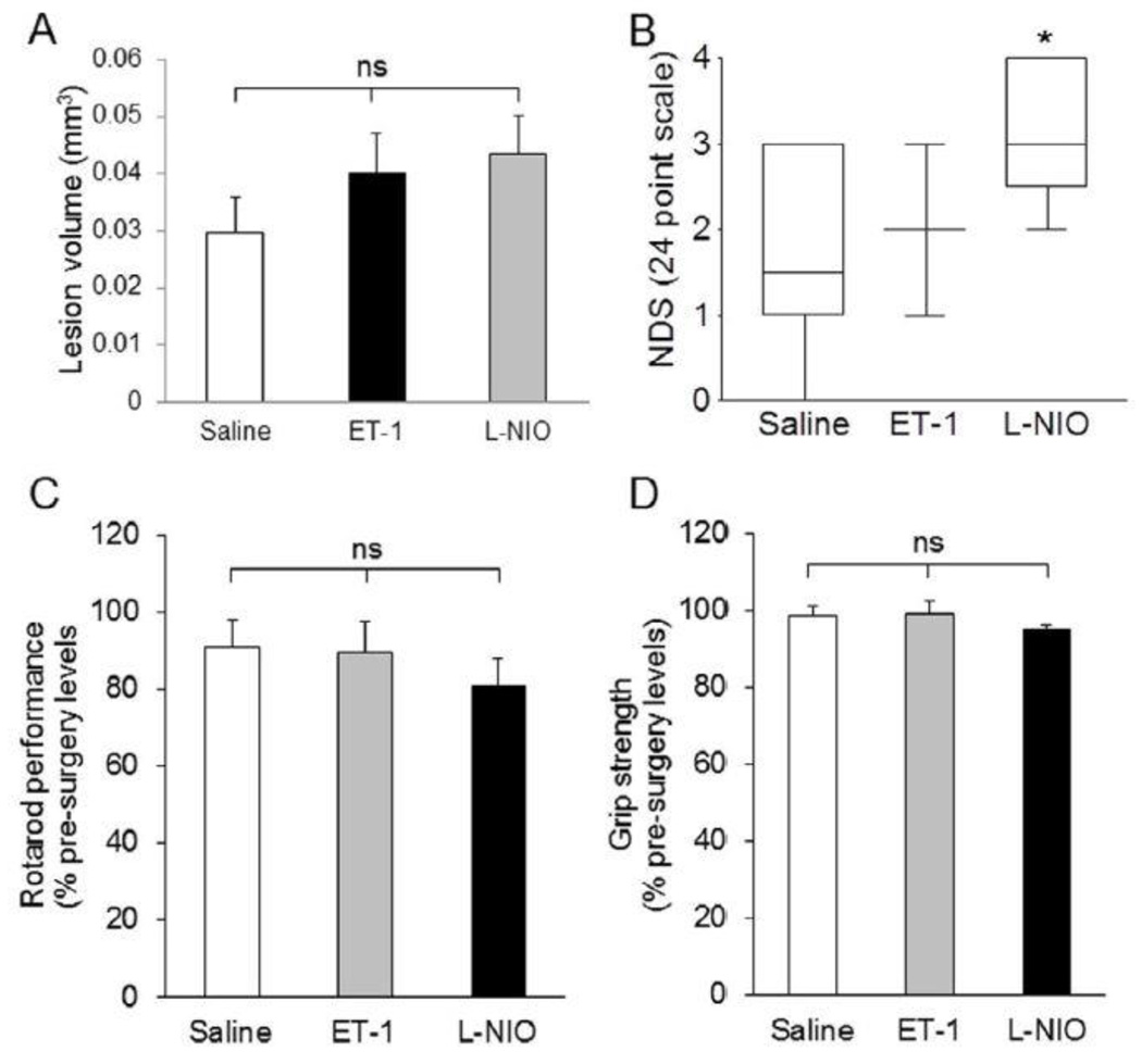
Figure 2.
Anatomical and functional outcomes after LPC, endothelin-1, or L-NIO injections in the PVWM. Although the lesion volume (A) in LPC-injected animals is higher compared with the saline-, ET-1, and L-NIO-treated mice, neurological deficit score was higher in L-NIO-injected groups (NDS; B). Other functional outcomes remained similar among the groups. Data presented as mean ± SEM. * P < 0.05, ** P < 0.01, when compared with the saline group; ns = non-significant.

