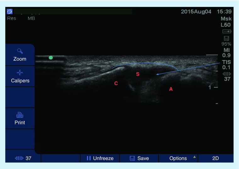
Figure 9. . Transverse sonogram of the acromioclavicular joint using a high-frequency linear probe.
The patient was in a seated position, the ipsilateral hand was placed on the contralateral shoulder. Arrow represents the projected path of needle. The arch represents the joint capsule.
A: Acromion; C: Clavicle; S: Joint space.

