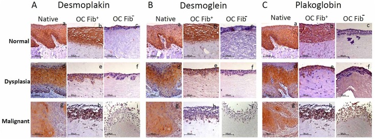
Fig 5. Immunohistochemical staining pattern of desmosomal proteins in in-vitro grown tissues.
IHC staining of desmoplakin (Aa-i), desmoglein (Ba-i) and plakoglobin (Ca-i) in Fib+ and Fib- OCs reconstructed from normal, dysplastic and malignant tongue tissues along with their respective native tissues. All three types of native tissues- normal, dysplastic and malignant showed similar immunolocalization and staining intensity for desmoplakin (Aa, b, d, e, g, h), desmoglein (Ba, b, d, e, g, h) and plakoglobin (Ca, b, d, e, g, h) in comparison with Fib+ OCs. While, Fib- OCs showed weak staining throughout all epithelial layers for desmoplakin (Ac, f, i), desmoglein (Bc, f, i) and plakoglobin (Cc, f, i). The experiments were performed at least three times.

