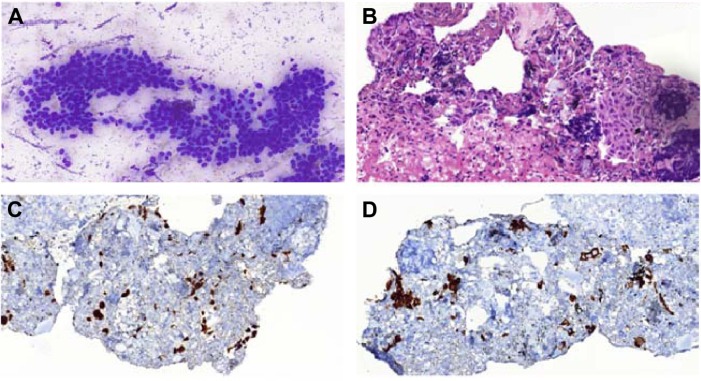
Figure 4.
Biopsy images.
Notes: (A) EBUS-FNA for mediastinal subcarinal lymph node: cytological appearance of nonkeratinizing squamous cell carcinoma. Sheets of atypical squamous cells with large hyperchromatic nucleus, dense squamoid cytoplasm, and moderate pleomorphism are observed (MGG stain, original magnification ×400). (B) A cell block section of tumor displaying few small atypical small squamous islands and scattered lymphocytes showing crushing artifact in a fibrinous background (H&E stain, original magnification ×400). Cell block immunohistochemistry of the tumor: tumor cells display strong nuclear p63 (C) and cytoplasmic cytokeratin 5/6 (D) positivities, which are characteristics for squamous cell carcinoma.
Abbreviations: EBUS-FNA, endobronchial ultrasound-guided transbronchial fine-needle aspiration; MGG, May–Grünwald–Giemsa; H&E, hematoxylin and eosin.

