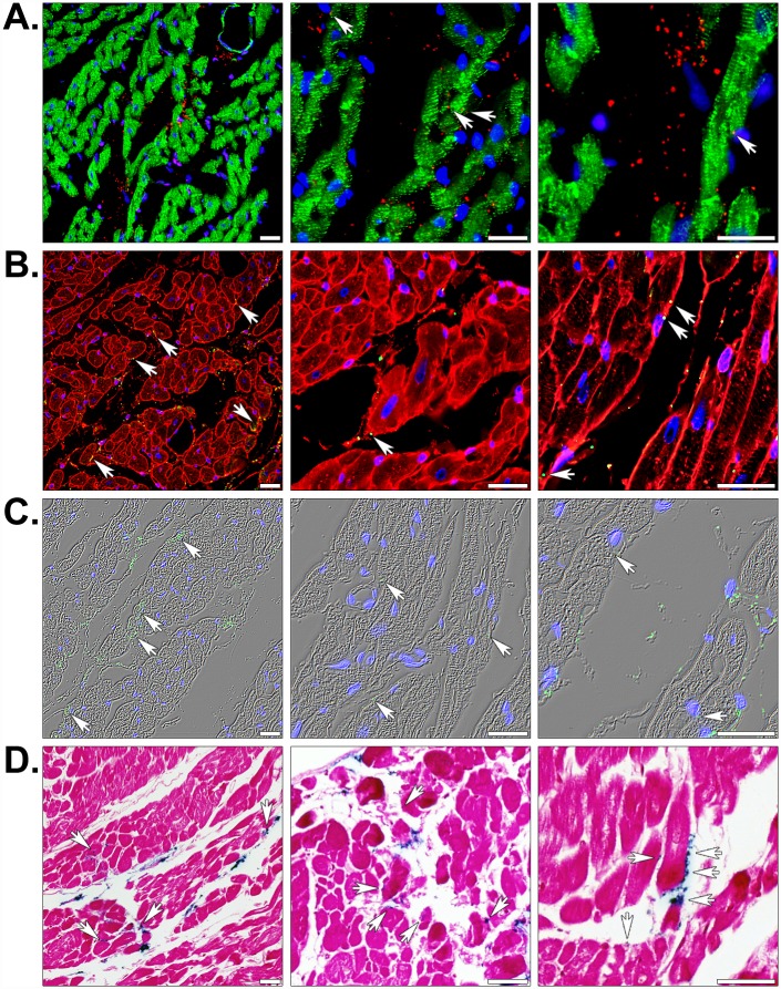
Fig 4. Histology of globally or regionally ischemic rabbit hearts injected with human mitochondria.
(A) Injected heart sections were fluorescently stained for the muscle markers desmin (green), the human-specific mitochondrial marker MTCO2 (red), and nuclei using the DNA stain DAPI (blue). (B) Fluorescent staining with the membrane marker WGA (red), the 113–1 human mitochondrial marker (green), and DAPI (blue). (C) MTCO2 and nuclear staining is shown with phase contrast illumination. (D) Prussian blue (blue) and pararosaniline (pink) staining of injected mitochondria labeled with magnetic iron oxide nanoparticles. Scale bars represent 25 μm. Transplanted mitochondria associated with cardiac myocyte sarcolemmata are indicated (arrows).

