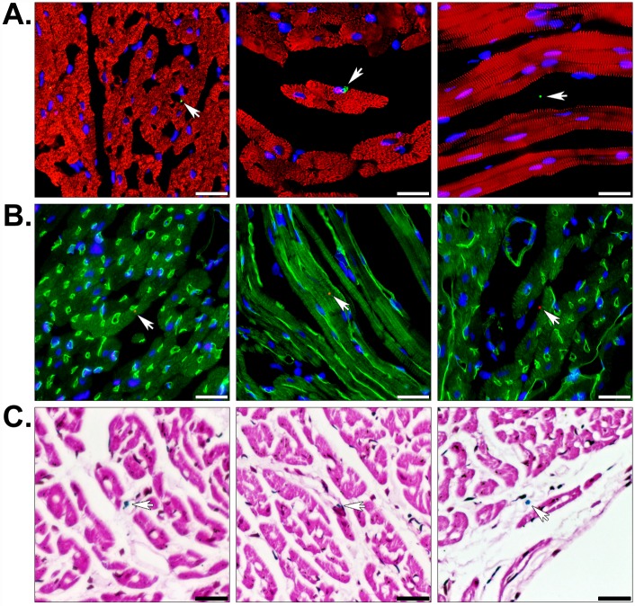
Fig 6. Histology of regionally ischemic hearts perfused with human mitochondria.
(A) Perfused heart sections were stained with the muscle marker α-actinin (red) and the human mitochondrial marker MTCO2 (green) to show the position of transplanted mitochondria in the heart (arrows). (B) Some hearts were also perfused with FITC-lectin (green) prior to fixation to display luminal surfaces of blood vessels. These sections were counter-stained with the 113–1 human mitochondrial marker (red) and nuclei were stained with DAPI (blue). The staining shows transplanted mitochondria associated with the vasculature, within interstitial spaces, and attached to cardiomyocytes. (C) Prussian blue staining (blue) and a pararosaniline counterstain (pink) confirmed transplanted mitochondria were labeled with magnetic iron oxide nanoparticles. Scale bars equal 25 μm.

