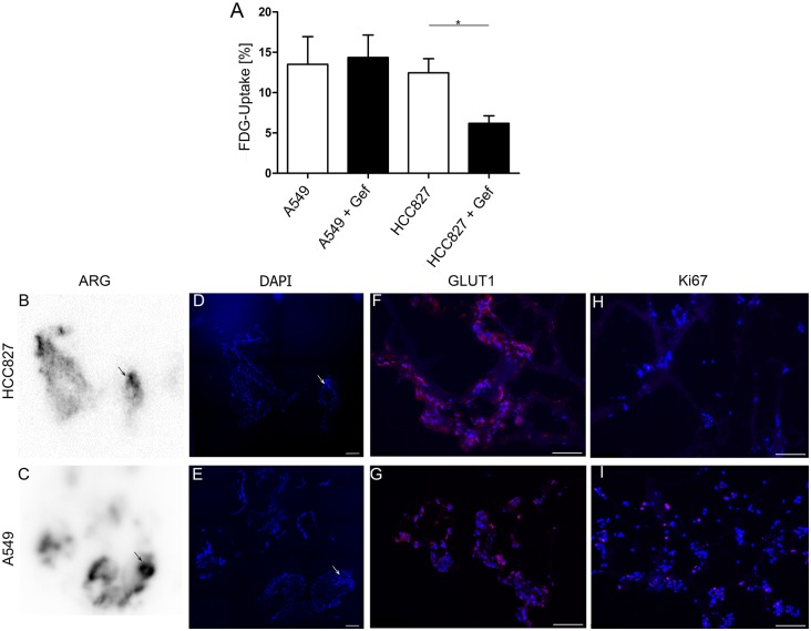
Fig 3. Detection of tumor nodules with FDG-PET.
(A) Gefitinib-treated and untreated A549 and HCC827 cells grown in 2D were incubated with 18F-FDG for 60 minutes and FDG uptake was quantified using a gamma-counter. Data were corrected for background and decay and related to the initially added activity (n = 4, p = 0.02, Man Whitney U test). (B-I) Cross sections of lungs recellularized with tumor cells and incubated with 18F-FDG were investigated by autoradiography (ARG). Regions of high radioactive intensity correlated with presence of tumor cells (arrows in B, C, D, E). Cells of both cell lines strongly expressed GLUT1 (F, G) while only a low amount of the tumor cells was proliferative as shown by Ki67 staining (H, I). Data are presented as arithmetic means ± SEM; *p<0.05, Man Whitney U test; scale bars: 50 μm. One representative experiment out of 3 is shown.

