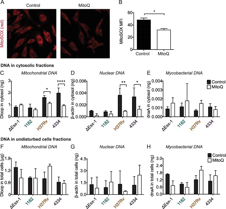
Fig 5. Mitochondrial stress contributes to accumulation of mtDNA in the cytosol during H37Rv/Lineage 4 and 4334/Lineage 2 infections.
A-H) BMDM were treated with MitoQ or control (dTPP) for 4 hours and then infected with the indicated mycobacterial strains at an MOI of 5. A) Uninfected cells were stained with MitoSOX at the time of infection. B) Mean fluorescence intensity (MFI) of MitoSOX was determined using ImageJ. C-H) 24 hr post infection, cells were collected and fractionated. Amount of DNA in cytosolic (C-E) and undisturbed cell (F-H) fractions was determined using gene-specific primers as in Fig 4A–4D; amount in ng or μg was determined using standards that were generated independently of experimental samples and that contained abundant levels of each gene. *p<0.05, **p<0.01, ****p<0.0001 by two-way ANOVA with Sidak post-tests; means ± SD (n = 3).

