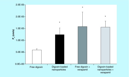
Figure 3. . Apparent permeability (Pe) for the transport of free digoxin (white bar) and digoxin-loaded polyethylene glycol–poly(lactic-co-glycolic acid) nanoparticles (RGPd50105, 10% drug loading, black bar) across BeWo cell monolayers in the apical (maternal) to basolateral (fetal) direction at the 2-h time point.
The transport studies were carried out at 37°C under cell culture conditions with stirring. Pe was also determined for both formulations in the presence of 100 μM verapamil, a P-gp inhibitor. Asterisks indicate significant differences from the permeability of free digoxin (p < 0.05). There were no significant differences between the Pe values of the digoxin-loaded nanoparticles, free digoxin in the presence of verapamil (dark gray bar) or digoxin-loaded nanoparticles in the presence of verapamil (light gray bar). Error bars indicate standard deviation (n = 3). PLGA: Poly(lactic-co-glycolic acid).

