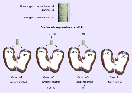
Figure 1. . A schematic representation of microsphere-based gradient scaffold and the various groups for implantation.
Gradient scaffolds were implanted into critical size osteochondral defects (D = 6 mm × H = 6 mm) in the lateral and medial femoral condyles of knee joints of group I animals. Group I–A received only the gradient scaffold (n = 6), and was the primary test group. Group I–B was a pilot group, where 1 µg TGF-β3 was added to the scaffold in the operating room immediately prior to implantation (n = 3). Similarly, 0.5 µg IGF-1 was added to the scaffold immediately prior to implantation in pilot group I–C (n = 3). Microfracture procedure was performed in group II animals (n = 6) following induction of full-thickness, chondral-only defects (D = 6 mm). The cartilage regeneration was evaluated 1 year post-implantation.
LFC: Lateral femoral condyle; MFC: Medial femoral condyle.

