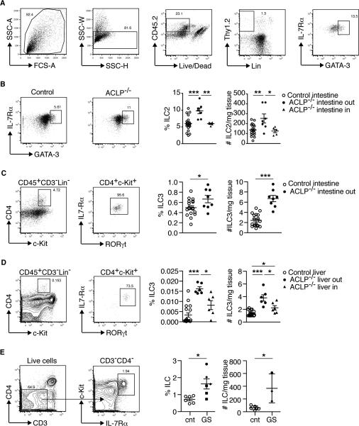
FIGURE 4.
Increased innate lymphoid cells (ILCs) in the exteriorized organs of ACLP−/− mice and patients with gastroschisis. (A) Sequential gating strategy to identify ILC2 (defined as CD45+Lin−CD3−Thy1.2+IL-7Rα+GATA-3+) in the intestine of a representative ACLP−/− mouse. (B) Representative flow cytometric plot of ILC2 and graphs showing the percentages and absolute cell number/mg tissue in the exposed (out) or non-exposed (in) intestine in littermate controls and ACLP−/− mice. Each symbol represents a single fetus; small horizontal bars indicate the mean. Control n=18, ACLP−/− n=12. *p<0.05; **p<0.01; ***p<0.001 by one-way ANOVA. (C and D) Gating strategy to identify ILC3 (defined as CD45+Lin−CD3−CD4+IL-7Rα+c-Kit+RORγt+) from E18.5 ACLP−/− mouse (example shown) and graphs showing the percentages (on total CD45+ cells) and absolute cell number/mg tissue in the exposed (out) or non-exposed (in) (C) intestine and (D) fetal liver in littermate controls and ACLP−/− mice. In this set of experiments, the entire intestine was exteriorized in the affected mice. Control n=6, ACLP−/− n=7. *p<0.05; **p<0.01; ***p<0.001 by one-way ANOVA. (E) Flow cytometric analysis of ILC (defined as CD3−CD4−IL-7Rα+c-Kit+) and compiled analysis of ILC of lamina propria and intraepithelial lymphocytes isolated from intestine of healthy controls (cnt, n=6 samples from 4 patients) and patients with gastroschisis (GS, n=6 samples from 3 patients). Among gastroschisis samples, each symbol (square, diamond and circle) represents a different patient; small horizontal bars indicate the mean. *p<0.05 by Mann-Whitney test.

