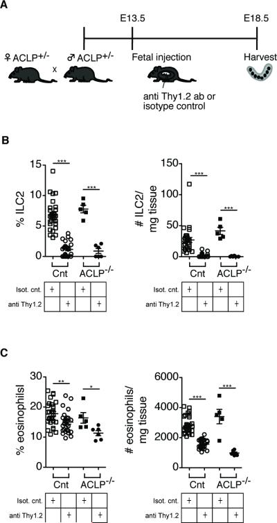
FIGURE 6.
Decreased eosinophil infiltration in ACLP−/− mice after fetal treatment with anti Thy1.2 antibody to deplete innate lymphoid cells (ILC). (A) Schematic layout of the experiment. Fetal mice are injected with anti Thy1.2 or isotype control antibody and harvested on E18.5. (B) Histograms showing the depletion of ILC2 (% and absolute cell number/mg tissue) in the intestine. (C) Histograms showing the reduction of eosinophils (% and absolute cell number/mg tissue) in the intestine. Each symbol represents a single fetus; small horizontal bars indicate the mean. Control n=56, ACLP−/− n=11. *p<0.05; **p<0.01; ***p<0.001 by Student's t test.

