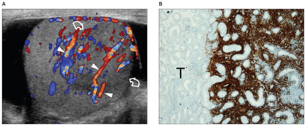
Figure 1.
Images from a 73-year-old patient with primary diffuse large B-cell lymphoma of the right testis presenting with a palpable testicular nodule. A, Color Doppler sonogram showing a unifocal hypoechoic lesion (curved arrows), which is hypervascular compared to the surrounding testis. Normal testicular vessels (arrowheads) cross the lesion with a rectilinear course, suggesting infiltrative disease. B, Immunohistochemical stain for CD20, a hallmark for B-cell lymphoma, showing the interface between the unstained uninvolved testis (T; left) and the portion of the testis involved by the tumor (right). CD20-positive tumor cells (brown) grow among the testicular vessels and seminiferous tubules without destroying them (original magnification ×20).

