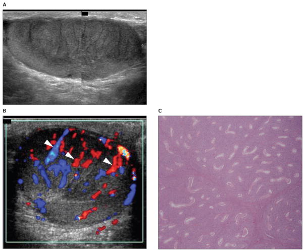
Figure 2.
Images from a 62-year-old patient with primary lymphoblastic lymphoma of the right testis presenting with a palpable right testicular nodule. A, Grayscale sonogram showing a striated appearance of a hypoechoic testicular mass involving most of the parenchyma. B, Color Doppler sonogram showing normal testicular vessels crossing the lesion (arrowheads). C, Photomicrograph of the histologic specimen showing diffuse peritubular and intratubular proliferation of lymphoma cells. Seminiferous tubules are infiltrated but not destroyed (hematoxylin-eosin, original magnification ×10).

