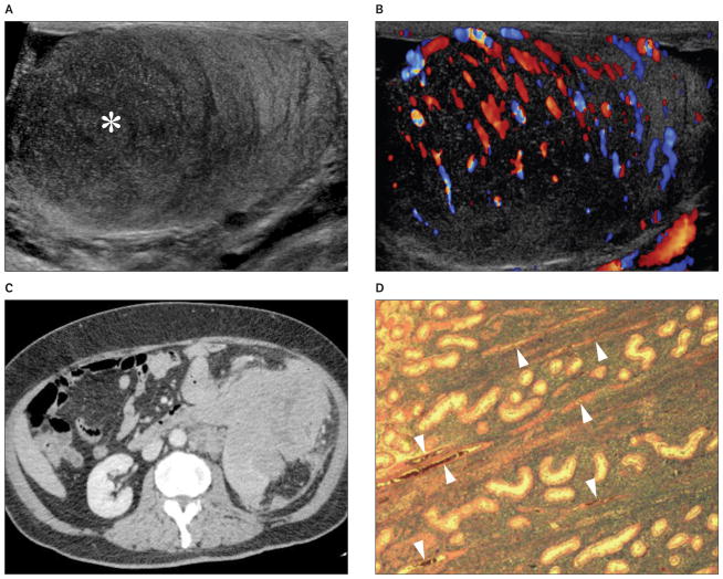
Figure 3.
Images from a 64-year-old patient with secondary lymphoma involving the left testis. The patient presented with a palpable left testicular nodule and abdominal mass. A and B, Grayscale (A) and color Doppler (B) sonograms showing a hypoechoic (asterisk) hypervascular lesion with poorly defined margins and normal intralesional testicular vessels. C, Contrast-enhanced computed tomogram showing extensive tumor involvement of the abdominal organs. D, Histologic specimen of the testis showing diffuse large B-cell lymphoma. Normal testicular vessels (arrowheads) and seminiferous tubules are surrounded by tumor cells (hematoxylin-eosin, original magnification ×20).

