. 2016 Jul 13;5:e15082. doi: 10.7554/eLife.15082

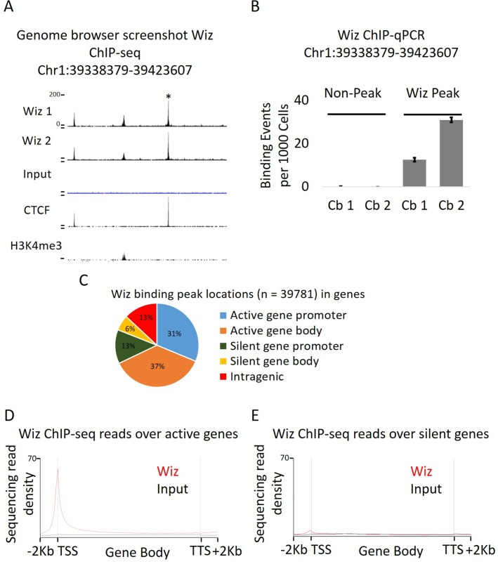
Figure 2. Wiz binds across the genome and at promoter elements.

(A) ChIP-seq was performed for Wiz in the adult male cerebellum, a screenshot is shown of a random Wiz-enriched site (* indicated) with the two ChIP-seq replicates and input, from the ~40,000 significantly enriched peaks. Encode data for CTCF and H3K4me3 is also included. (B) Enrichment for Wiz in two cerebellum samples (Cb1 and Cb2) is shown by ChIP-qPCR, with primers located in regions not enriched for Wiz in ChIP-seq data and primers flanking the * indicated ChIP-seq peak from (A). Enrichment is represented as binding events detected per 1000 cells and are generated by running samples in parallel with known amounts of genomic DNA. Error bars indicate S.D. from 3 technical replicates. (C) The percentage is shown of Wiz peaks that overlap with the promoter (up to 2 kb from a TSS) and genic sequence of genes that were classified as active or silenced, according to RNA-seq data mapped to Ensembl genes annotations. (D) Wiz ChIP-seq (and Input) occupancy over all Ensembl gene bodies is shown as deep sequencing read density along the transcription unit, including 2 kb up and downstream of the transcriptional start and stop site. Genes were separated into either active or silenced transcriptional states, as in C.

DOI: http://dx.doi.org/10.7554/eLife.15082.003

Figure 2—source data 1. Anti-Wiz antibody co-immunoprecipitation.
(A) Nuclear lysates from either pooled E13.5 embryonic brain or 2 adult cerebellum replicates were subjected to antibody-based affinity purification and mass spectroscopy. The number of peptides are shown for each protein identified in both embryonic (values for 1 sample) and adult brain (values for each of the two samples) anti-Wiz antibody IP samples and absent in anti-IgG antibody controls run for each sample, with greater than 5 peptides in each IP. Proteins are ranked by the semi-quantitative number peptides in embryonic brain where Wiz expression is higher. The previously identified Wiz-Zfp644-EHMT1-EHMT2 complex is shaded.
DOI: 10.7554/eLife.15082.004

Figure 2.

Figure 2—figure supplement 1. Anti-Wiz antibody western blotting.
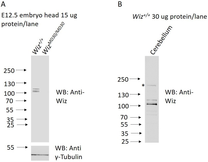
Figure 2—figure supplement 1.

(A) Western blotting for Wiz using an NBP180586 anti-Wiz antibody is shown for total protein lysates from E12.5 Wiz+/+ and WizMommeD30/MommeD30 embryo head. Three bands at ~100 kD represent Wiz isoforms, consistent with UCSC gene splice variants, in Wiz+/+ sample and are absent in WizMommeD30/ MommeD30 sample. Western blotting with an anti-γ-Tubulin antibody is shown as a loading control. (B) Anti-Wiz antibody staining in adult cerebellum, showing the short (~100–120 KDa) isoforms present in embryo head protein extract, as well as a long (~160 KDa) isoform previously reported to be expressed (Matsumoto et al., 1998).