. 2016 Jul 19;5:e17262. doi: 10.7554/eLife.17262

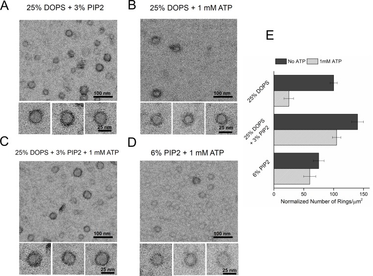
Figure 3. Syt1-PIP2 interaction is key to ring-formation under physiologically relevant conditions.

(A) Inclusion of 3% PIP2 (in addition to 25% PS) in the monolayer stabilized the ring structures and increased the number of rings observed. (B, C) Addition of ATP drastically reduced the number of rings observed in monolayers containing 25% PS only, but not when supplemented with 3% PIP2. (D) PIP2 (6%) as the only anionic lipid on the bilayer was sufficient to assemble ring-like oligomers, even in the presence of 1 mM ATP. All EM analyses were carried out using 5 µM protein in buffer containing 100 mM KCl and 1 mM free Mg2+. Representative micrographs and average values/SEM from a minimum of three independent trials are shown in (E). The rings observed under all conditions shown in (E) were similarly (~30 nm) sized

DOI: http://dx.doi.org/10.7554/eLife.17262.009

Figure 3.

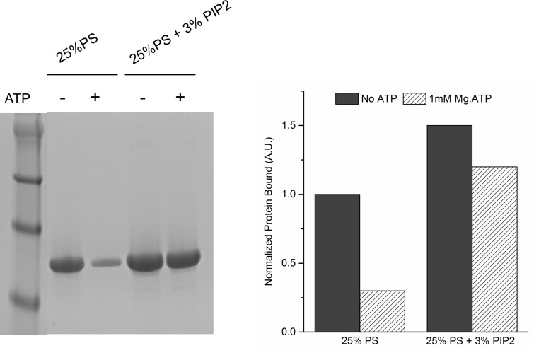
Figure 3—figure supplement 1. Lipid binding analysis shows that ATP effectively screens the interaction of Syt1CD to PS-only membrane, but not membrane containing 3% PIP2.

Figure 3—figure supplement 1.

To assess Syt1CD-membrane binding, 10 µM of Syt1CD were mixed with 1 mM small unilamellar vesicles (SUV) containing either 25% PS or 25% PS+3% PIP2 (remainder was PC) and incubated for 1 hr at RT with in in buffer containing 100 mM KCl and 1 mM free Mg2+ supplemented with 1 mM Mg2+ or 1 mM Mg-ATP. The SUVs were isolated using discontinuous density gradient and analyzed on SDS-PAGE/ Coomaisse analysis after adjusting for the amount of lipid recovered. The amount of protein bound in each case was estimated using density measurement using ImageJ software.