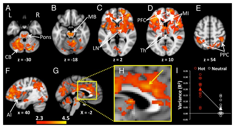
Figure 2. Increased levels of functional connectivity with the POA during heating compared with the neutral condition were seen in widely distributed regions of the brain. (A) The pons and bilateral regions of the cerebellum (CB) showed heating-related increases of POA connectivity. (B) Enhanced POA connectivity was also apparent in midbrain (MB), (C) lentiform nuclei (LN), (D) bilateral thalamus (Th), mid insula (MI), prefrontal cortices (PFC), (E) and bilateral posterior parietal cortices (PPC). (F) The anterior insula (AI) in the right hemisphere showed heating-related increases in connectivity with the POA. (G) Mesial regions including the midcingulate cortex, posterior cingulate cortex and precuneus had increased POA connectivity during heating. (H) An enlarged image of the mesial surface of the brain shows the location of increased POA connectivity in the midcingulate cortex The arrow indicates the voxel with the highest statistic (z = 3.73) in the slice at x = –2, y = 18, z = 28. (I) Signals from the midcingulate region, shown in H, were extracted and correlated with the corresponding POA seeds in order to visualize the distribution of variances between the two groups of participants. Activations Pcorrected < 0.05. Background image is MNI template brain and axial slices are oriented to show right side of brain on right side of image. Positions of slices labeled according to convention where +ve z = distance in mm superior to anterior commissure, -ve z = distance in mm inferior to anterior commissure, +ve x = distance in mm to right of midline, -ve x = distance in mm to left of the midline. Axial images (A–E) are displayed with the right side of the brain on the right side of the image.

An official website of the United States government
Here's how you know
Official websites use .gov
A
.gov website belongs to an official
government organization in the United States.
Secure .gov websites use HTTPS
A lock (
) or https:// means you've safely
connected to the .gov website. Share sensitive
information only on official, secure websites.
