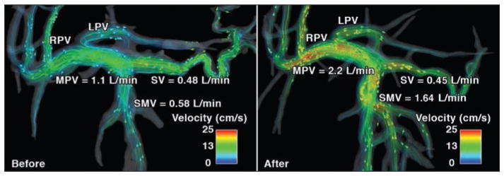
Fig. 3.
32-year-old man (79.5 kg) with no history of liver disease (case 2). Velocity distribution shown by velocity color-coded streamlines on 4D flow MR images obtained before (left) and after (right) meal challenge reveals blood flow increase in superior mesenteric vein (SMV), left portal vein (LPV), right portal vein (RPV), and main portal vein (MPV) in response to meal challenge. Arrowheads show direction of blood flow. Reduction in splenic vein (SV) flow can also be observed.

