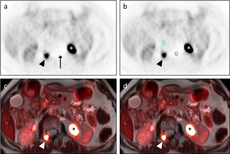
Fig. 1.
Outlier FFBL resulting from scatter correction artifact. A 69-year-old man with metastatic papillary thyroid carcinoma status-post multiple radioiodine treatments presented for restaging due to a rising thyroglobulin. Transaxial PET images (a, b) and transaxial T2W HASTE images with PET fusion (c, d) demonstrate an FFBL within the right posterior elements of the L1 vertebra (arrowhead) as well as significant tracer activity within a dilated left renal collecting system (asterisk). The initial BB contour (pink circle in b, d) for this FFBL was drawn in the left posterior elements of the L1 vertebra. Statistical analysis showed this lesion to be an outlier in terms of the ratio of FFBL SUV-mean to BB SUV-mean for both the PET/CT and PET/MRI datasets. SUV-mean for this initial BB contour was 0.39. While not readily apparent on the fusion images, inspection of the PET images (a, b) revealed an area of photopenia (arrow in a) in which this contour (pink circle in b) had been placed. This photopenia was consistent with scatter correction artifact from the excreted FDG in the adjacent left renal collecting system (asterisk). Moving the BB contour out of this photopenic region to the right L1 vertebral body (blue circle in b, d) for both the PET/CT (not shown) and PET/MRI datasets resulted in a new BB SUV-mean of 1.1, similar to values obtained at other vertebral levels. Repeat statistical analysis showed no outliers

