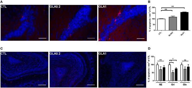
Figure 5.
Apoptotic cells labeling in SVZ and in the olfactory bulb. Apoptag® immunostaining of coronal sections of CTL (n = 3), GLA0.2 (n = 3) or GLA1 (n = 3) in the SVZ (A) and in the OB (C) showing a dose effect of GLA in the increase of apoptotic cells in the SVZ of exposed mice (B). Unlike to SVZ, we show a significant decrease of the number of apoptotic cells in the granular layer (Grl) of exposed mice (D). No difference was found in neuro-epithelium (NE) and in glomerular layer (Glo). Each value represents the mean ± SEM (*p < 0.05, ***p < 0.001). Scale bar 100 μm.

