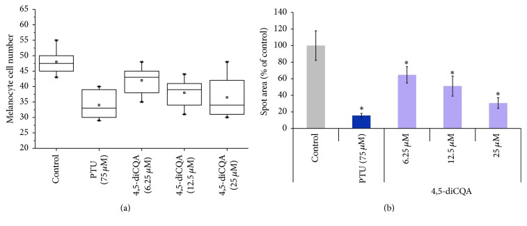
Figure 6.
The effect of 4,5-diCQA on melanocyte cell number and spot area in zebrafish embryos. (a) Box and whisker plots of melanocytes number in the head region of 4,5-diCQA-treated embryos indicated no major difference compared with untreated embryos. The standard mean is indicated; outliers are represented by an asterisk. (b) Surface area of the melanocytes in zebrafish embryos. PTU was used as positive control. Azelaic acid (AZ) was used as a positive control. The results are expressed as percentages of control, and the data are the means ± SEM of three independent experiments. ∗ P < 0.05 as compared to the untreated control.

