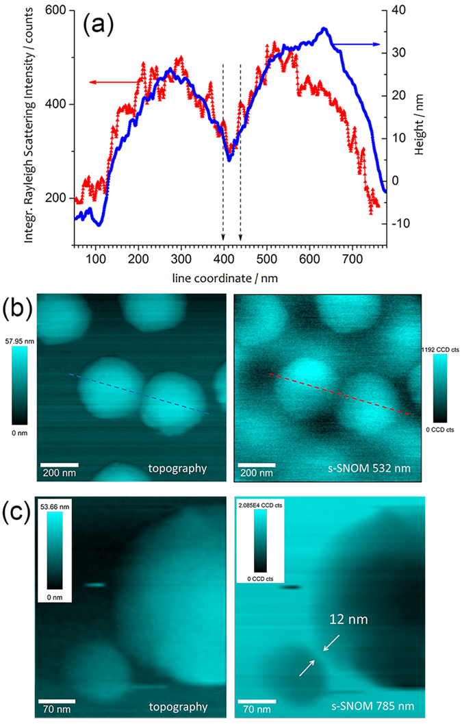
Figure 8. s-SNOM measurements on a patterned gold nanostructure.

(a) Topography and integrated Rayleigh intensity (s-SNOM) along the cross section indicated in (b), acquired in contact mode with a Ag@AuNP coated tip, with incident power of 100 nW at 532 nm and integration time of 15 ms per pixel, with incident radial polarization, over an area of 1 μm2 (256 × 256 pixels). (c) Detail of closely spaced gold nanopillars (topography and s-SNOM), extracted from a scan area of 350 × 350 pixels over 4 μm2 (step of 5.7 nm), measured with excitation laser power of 150 nW at 785 nm, with radial polarization and integration time of 12 ms per pixel. All colormaps represent signal amplitudes leveled to the minimum.
