Figure 5.
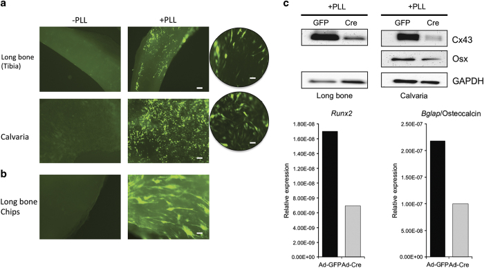
PLL can be used to drastically enhance transduction of osteogenic cells in situ. (a) Fluorescent images of intact tibias and calvaria transduced with Ad-GFP in the absence (left side) or presence (right side) of PLL. Scale bar equals 100 μm. A higher magnification view (circles) is also displayed. Scale bar equals 20 μm (b) Smaller long bone fragments transduced with Ad-GFP in the absence (left) or presence (right) of PLL imaged. Scale bar equals 20 μm. (c) Western blot probing for Cx43, Osterix (calvaria only) and GAPDH using tissue homogenates obtained from long bone pieces and calvaria transduced with Ad-GFP and Ad-Cre in the presence of PLL only. Quantitative real time RT-PCR of the mRNA levels of Runx2 and Bglap/osteocalcin from the adenoviral transduced calvaria shown in (a). GFP, green fluorescence protein; MOI, multiplicities of infection; PLL, poly-l-lysine; RT-PCR, reverse transcription–PCR.

