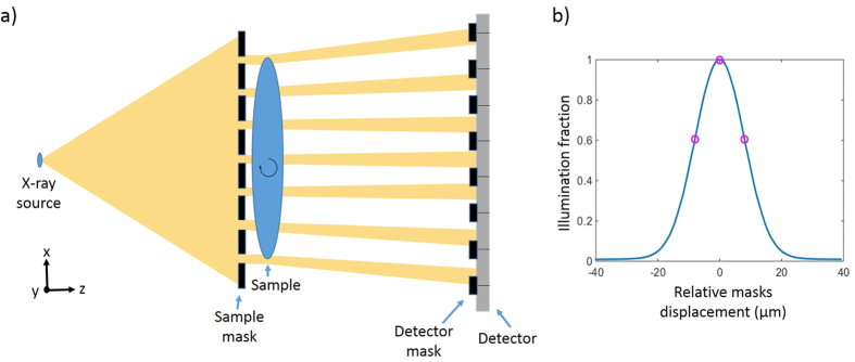
Figure 1.
(a) A top view of an EI setup with an extended x-ray source. For CT, the axis of rotation is aligned with the y direction, while the sample is moved by sub-pixel steps along x for dithering. (b) A typical illumination curve showing intensity variation as a function of masks displacement over one period. The circles represent regular choices of mask positions for imaging.

