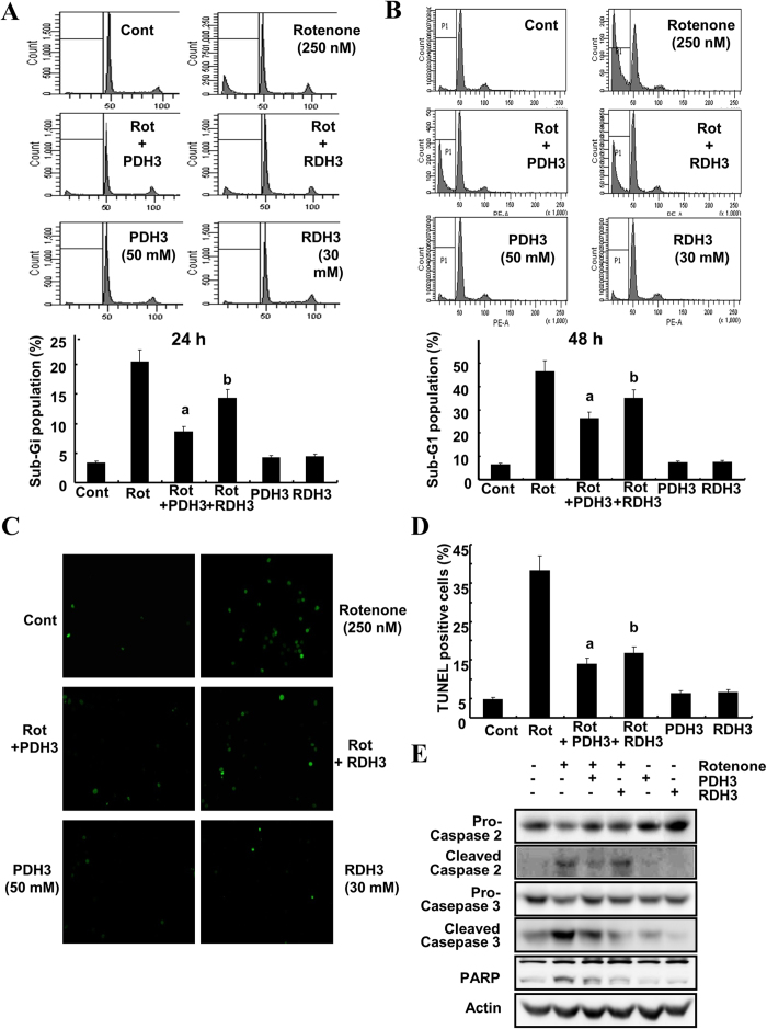
Figure 6. RDH3 and PDH3 partly inhibit apoptosis induced by rotenone treatment of SH-SY5Y cells.
(A,B) RDH3 and PDH3 partly inhibit apoptosis induced by rotenone treatment in SH-SY5Y cells. After TAT fused peptides were pre-incubated for 1 hour, the cells were treated with rotenone. After 24 h (A) or 48 h (B), apoptosis was analyzed as a sub-G1 fraction by FACS (Top; FACS histogram, Bottom; graph of sub-G1 population). Data shown are the means ± SD (n = 3). a, bp < 0.05 for rotenone-treated cells versus rotenone + PDH3-, or rotenone + RDH3-treated cells by ANOVA. (C,D) Rotenone-induced DNA fragmentation was reduced by RDH3 and PDH3. DNA fragmentation was measured by TUNEL staining of SH-SY5Y cells. Representative pictures show TUNEL staining (C). A DNA fragmentation detection kit was used to measure the fragmented DNA (D). (E) RDH3 and PDH3 partly suppress cleavage of procaspase-2 induced by rotenone treatment in SH-SY5Y cells. After TAT fused peptides were pre-incubated for 1 hour, the cells were treated with rotenone for 48 h. Equal amounts of cell lysates (40 μg) were subjected to electrophoresis and analyzed by Western blot for caspase-2, caspase-3, and PARP. Actin was used for normalization.

