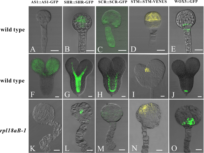
Figure 5. Cell-type specific expression pattern of marker genes were disrupted in rpl18aB-1/rpl18aB-1 embryos.
The upper arrow showed the globular embryos isolated from different marker lines. Both normal ovules and abnormal ovules in the same silique of different marker lines in rpl18aB-1 could be found. The middle arrow showed isolated heart-stage embryos in normal ovules and the under arrow showed isolated rpl18aB-1/rpl18aB-1 embryos in abnormal ovules.(A–C) AS1::AS1-GFP expression in wild-type globular embryo (A), heart-stage embryos (B) and rpl18aB-1/rpl18aB-1 embryos (C). (D–F) SHR::SHR-GFP expression in wild-type globular embryo (D), heart-stage embryos (E) and rpl18aB-1/rpl18aB-1 embryos (F). (G–I) SCR::SCR-GFP expression in wild-type globular embryo (G), heart-stage embryos (H) and rpl18aB-1/rpl18aB-1 embryos (I). (J–L) STM::STM-VENUS expression in wild-type globular embryo (J), heart-stage embryos (K) and rpl18aB-1/rpl18aB-1 embryos (L). (M–O) WOX5::GFP expression in wild-type globular embryo (M), heart-stage embryos (N) and rpl18aB-1/rpl18aB-1 embryos (O). Bars = 20 μm for (A) to (O).

