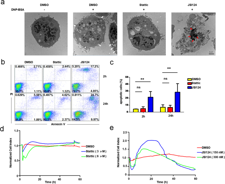
Figure 5. JSI124 induces apoptosis in RBL-2H3 cells.
(a) Mast cells treated with JSI124 exhibited the characteristics of apoptosis. RBL-2H3 cells were treated with DMSO, Stattic (6 μM) or JSI124 (300 nM) for 2 hours and stimulated with DNP-BSA for 15 minutes before imaging by transmission electron microscope (TEM). Representative images of cells are shown. White arrows indicate membrane fusion; Red arrows indicate chromatin condensation. (b,c) Annexin V and PI double staining assay illustrated that JSI124 promoted the apoptosis of mast cells. (b) RBL-2H3 cells were treated with inhibitors for 2 hours or 24 hours. Cells were stained with FITC-labeled annexin V and PI and analyzed by flow cytometry as described in Materials and Methods. (c) The percentage of apoptotic cells was calculated as follows: 100% - the percentage of double negative cells. (d,e) TCRP of JSI124 was apoptosis-related. RBL-2H3 cells were seeded in the plate for 24 hours, and the medium was subsequently replaced with serum-free fresh DMEM. After the addition of inhibitors, a typical dynamic cell response curve was recorded to detect cell apoptosis.

