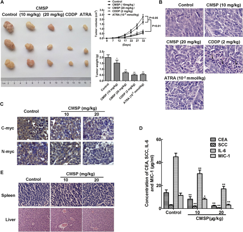
Figure 6. Inhibition of Kyse30 cell growth in vivo by treatment with CMSP.
(A) CMSP treatment inhibited tumour growth of Kyse30 cells in vivo. Left: Representative images of tumours formed in nude mice. Right: Tumour growth curves of xenograft tumours and measurement of the tumour weight (n = 6 for each group). (B) Histological analysis of the tumour tissues of mice in all of the groups. Representative H&E images are shown (×400). (C) Representative immunohistochemical images showing the expression levels of C-myc and N-myc in the tumour tissues of both groups (×400). (D) The content of CEA, SCC, IL-6 and MIC-1 in the serum of nude mice treated with CMSP was assayed by ELISA. The data presented are means ± SD from at least three independent experiments. **P < 0.01, compared with the control group. (E) Histological analysis of sections from the spleen and livers of mice treated with CMSP or untreated mice. Representative H&E images are shown (×400).

