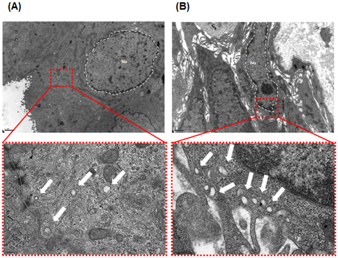
Figure 1. Representative transmission electron miscroscopy (TEM) images of extracellular vesicles (EVs) in prostate tissue.
Vesicles 30–100 nm in diameter were observed by TEM. (A) Human benign prostatic hyperplasia (BPH) cells produce several microvesicles. The lower panel shows a magnified region of (A). The EVs appear as white dots (indicated by an arrow). (B) Human prostate cancer cells shed more microvesicles compared to BPH cells. The lower panel shows a magnified region of (B) Bars in low-magnification images, 1 μm. Bars in high-magnification images, 200 nm.

