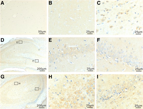
Fig. 1.

Immunohistochemical labeling of AhR in neurons of normal brains (b, d, e, and f) and brains 3 days post-TBI. a The specificity of AhR antibody was demonstrated by staining without the primary antibody and no IR was seen. b, c Examples of coronal sections of the cortex from normal brains (b) and brains 3 days after TBI (c). AhR expression (blue dots) was observed in the neurons of brain cortex in normal brains (b) and elevated three days post injury (c). d–i Examples of coronal sections of the AhR expression in hippocampus from normal brains (d–f) and brains 3 days after TBI (g–i). AhR expression was observed in neurons of pyramid cell layer of CA fields (e and h) and granular layer of dentate gyrus (f and i) in normal brains (d–f) and elevated in brains 3 days after TBI (g–i). The boxed areas indicate regions further observed under high-power magnification. The lower boxed areas represent the area of dentate gyrus and the upper boxed areas hippocampal CA fields
