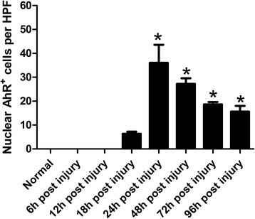
Fig. 2.

Time course of lesional AhR+ non-neuron cell accumulation in TBI. The numbers of parenchymal AhR+ non-neuron cells of every rat brain coronal section were counted in 8 HPFs. In each field, only positive cells with the nucleus at the focal plane were counted. Results were presented as arithmetic means of positive cells per HPF and standard errors of means (SEM). Statistical analysis was performed by one-way ANOVA followed by Dunnett’s Multiple Comparison test. *: p < 0.05 compared to normal control
