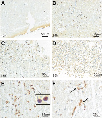
Fig. 3.

Accumulation of AhR+ non-neuronal cells in traumatic brains. In 12 h brains, AhR+ non-neuronal cells were rarely seen (a). Accumulation of AhR+ non-neuronal cells in the lesioned regions at day 1 (b), 2 (c) and 4 (d), respectively, after TBI. e Micrographs show that most AhR+ non-neuronal cells exhibited lymphocyte morphology with small cell body and cytoplasm (blue and brown dots). f Occasionally, the localization of AhR+ lymphocyte-like cells in the peri-vascular spaces corresponding to peri-vascular spaces was observed, indicating the blood origin. Furthermore, in AHR+ non-neuron cells, AhR was mainly localized to nucleus, suggesting an activated status (e and f)
