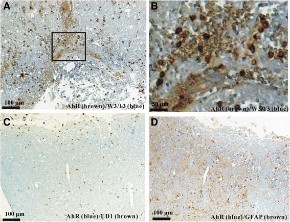
Fig. 4.

AhR double labeling in brain sections from day 1 after TBI. a Most AhR+ non-neuronal cells (brown) co-expressed W3/13 (blue). The boxed areas indicate the regions that were further observed under high-power magnification shown in (b). c and d: However, most AhR+ non-neuronal cells (blue) did not co-localize with ED1+ microglia (brown, c) or GFAP+ astrocytes (brown, d). Scale bar = 100 μm for (a, c) and d; 50 μm for (b)
