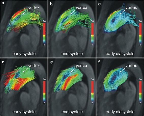
Fig. 2.

Vortical blood flow in the main pulmonary artery. Velocity-color-encoded streamlines (a, b, c) and 3D velocity vectors (d, e, f) projected onto multi-planar reformatted anatomical images demonstrate counter-clockwise rotating vortical blood flow nested in bi-directional pulmonary flow caused by PDA. This structure is present throughout the entire cardiac cycle (Additional file 2: in the online-only Data Supplement)
