
Figure 2.
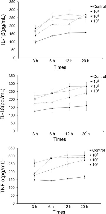
Establishing a model of infection of porcine peripheral blood monocytes by H. parasuis. 5 × 10⁵ cells were seeded into the culture plates, then H. parasuis (105, 106, 107 CFU/mL) was added to each well and incubated under 5% CO2 at 37 °C for 3, 6, 12, and 20 h, respectively. Inflammatory cytokines from the supernatant were measured to determine the MOI and optimal interaction time.