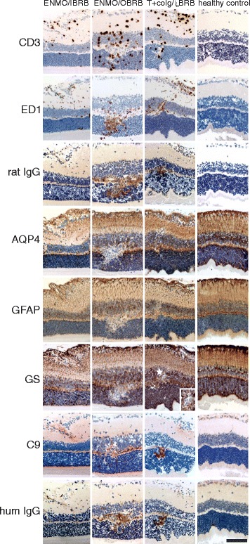
Fig. 5.

Histological characterization of inflammatory lesions in the retina. Consecutive retinal sections were reacted with antibodies against CD3, ED1, rat IgG, AQP4, GFAP, glutamine synthetase (GS), C9, and human IgG. Positive reaction products are shown in brown or red (C9 only). All sections were counterstained with hematoxylin to reveal nuclei in blue. Bars = 100 μm. The tissue derived from animals injected with AQP4268–285-specific T cells and NMO-IgG (ENMO), with AQP4268–285-specific T cells and human control IgG (T + co IgG) and healthy controls, and show inflammation at the inner blood retinal barrier (IBRB), at the outer blood retinal barrier (OBRB), simultaneously at both retinal barriers (I/OBRB), or an absence of inflammation at these sites in the control animals. The insert shown for the GS stained retina of the rat injected with T cells and co IgG refers to the area labeled by a white star in the adjacent overview. It demonstrates that there is no loss of GS reactivity at this site, but the lumen of a blood vessel. Bar = 100 μm
