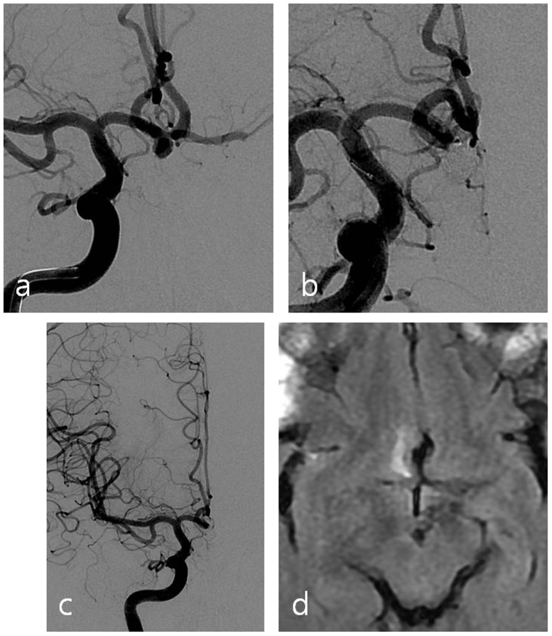
Figure 1.
(a) Working position angiography shows an aneurysm with a wide neck located at the anterior communicating artery. (b) Aneurysm and anterior communicating artery are both embolized with coils. (c) Angiography with left internal carotid artery compression after the procedure reveals occlusion of the anterior communicating artery. (d) Follow-up brain magnetic resonance imaging (MRI) shows infarction in right basal forebrain.

