Figure 9.
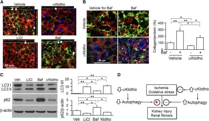
αKlotho-induced reduction of Col I accumulation is associated with increase in autophagic flux in OK cells. OK cells were seeded on coverslips and transfected with GFP-Col I plasmid. After 24 hours of transfection, cells were treated with autophagy inducer (LiCl; 10 mM) or suppressor (Baf; 200 nM) or αKlotho (0.4 nM) or vehicle. Cells were stained with anti-αKlotho and examined by confocal fluorescent microscopy. Rhodamine-phalloidin served as the counterstain. (A) Representative immunocytochemistry for αKlotho (blue), Col I (green), and phalloidin (red) in OK cells analyzed by laser confocal microscopy. Intracellular collagen is depicted by arrows, and extracellular collagen accumulation is depicted by arrowheads. Scale bar, 50 μm. (B, left panel) Representative immunocytochemistry for αKlotho (blue), Col I (green), and phalloidin (red) in OK cells. Scale bar, 50 μm. (B, right panel) Collagen in OK cells stained and quantified with the Sirius Red/Fast Green Kit. (C) OK cells were treated with LiCl (10 mM), Baf (200 nM), and αKlotho (0.4 nM) versus vehicle for 24 hours, and lysates were immunoblotted. (Left panel) Representative immunoblots for LC3-I and LC3-II, p62, and β-actin. (Right panel) Summary of immunoblots from three independent experiments. Data are expressed as means±SDs, and statistical significance was assessed by one-way ANOVA followed by Newman–Keuls test. Veh, vehicle. Statistical significance was accepted when *P<0.05; **P<0.01 between two groups. (D) Proposed working model of the effect of αKlotho on autophagy in AKI. αKlotho maintains a normal level of autophagy. Low αKlotho reduces and high αKlotho increases autophagic flux. Upregulation of autophagy exerts both cytoprotective and antifibrotic actions. In addition, αKlotho also protects the kidney against injury from oxidative stress or ischemia through antioxidation and antiapoptosis, which are independent of regulation of autophagy.

