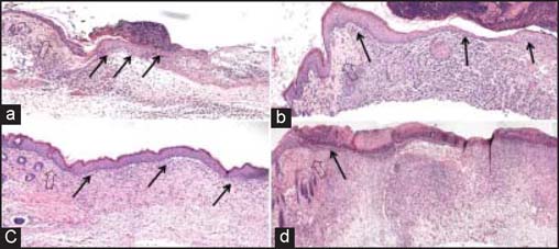
Figure 6:

Histological analysis of wound healing following CP and CPA grafting. Accelerated keratinocyte migration and re-epithelialization was seen in CPA-grafted mice. (a) CPA at day 5, (b) CPA at day 7, (c) CPA at day 14, and (d) CP at day 14. Each large arrow points to wound edge, while the small black arrows highlight keratinocyte migration and epidermal development.
